Hip And Leg Bone Diagram : Hip Fracture Cedars Sinai : Start studying leg bone diagram.. Leg bones anatomy, function & diagram | … 06.08.2020 · hip pain location diagram. When you stand or walk, all the weight of your upper body rests on them. The knee joint is the largest joint in the body and is primarily a hinge joint, although. The head of your femur fits into your hip socket and the bottom end connects to your knee. The foot bones shown in this diagram are the talus, navicular, cuneiform, cuboid, metatarsals and calcaneus.
Learn about hip and leg bones with free interactive flashcards. When the leg is stretched out, the knee joint is placed on a straight line with the hip and ankle (left). Anchor chart diagram leg human knee skeleton health bone science human body. Bringing the leg back towards the midline. The ilium bone forms the superior portion of the os coxa, the ischium bone the lower posterior portion, and the pubic bone (pubis) the lower anterior portion.
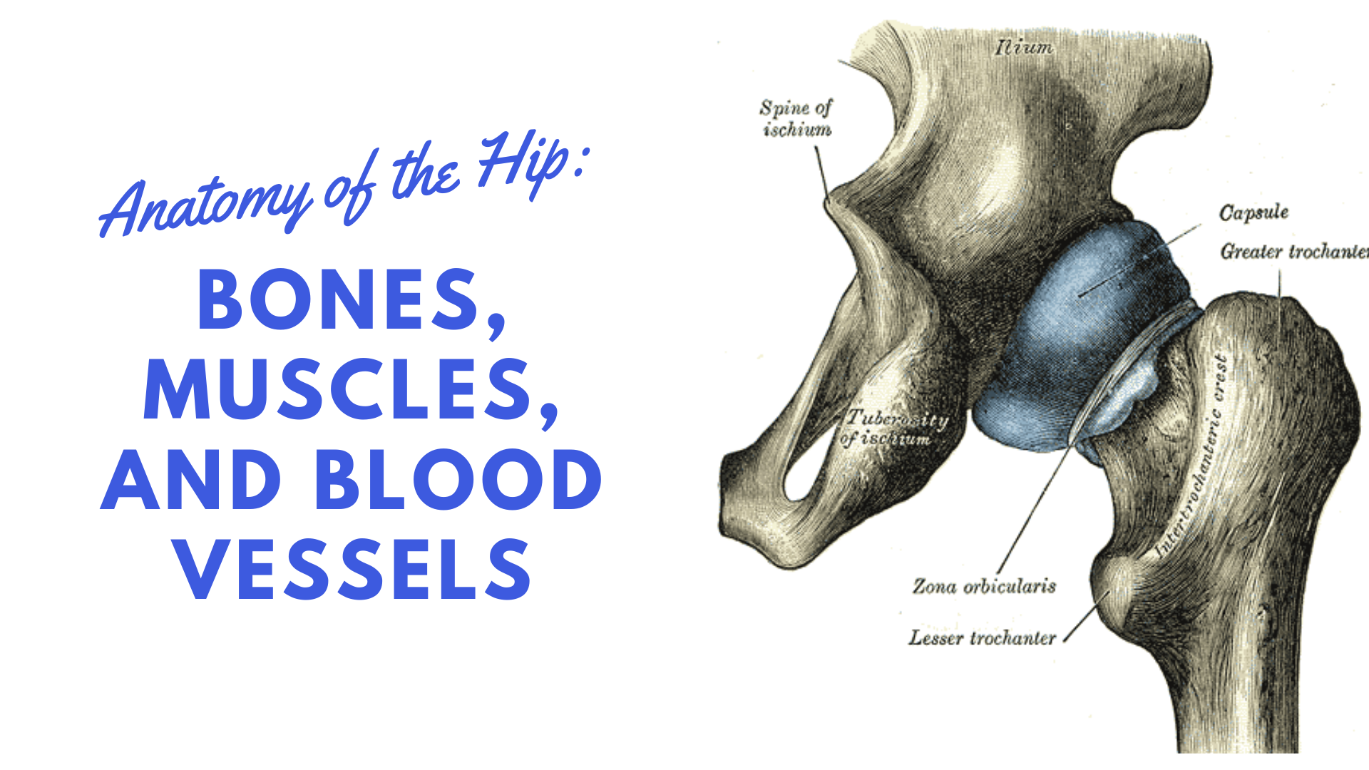
Clinical Anatomy The Bones Of The Knee And Leg Dummies from www.dummies.com In some vertebrates (including humans before puberty) it is composed of three parts: This bone attaches to the sacrum (forming the sacroiliac joint) and to its counterpart at the pubic symphysis, forming the pelvic girdle. These muscles include the adductors (adductor magnus. Click and start learning now! The ilium, ischium, and the pubis. The bone surfaces of the femoral head and acetabulum have a smooth durable layer of articular cartilage that cushions the ends of the bones and allows for smooth movement. It joins the lower limb to the pelvic girdle. Bones of the hip joint.
Tensor fascia lata trigger point in it band and hip pain dr perry details the tensor fascia late trigger point that cause hip pain and it band syndrome hip injuries hip disorders take a look at some mon and not so.
Bones of the hip joint. Spine bones diagram unique simple bone. These muscles include the adductors (adductor magnus. These same nerves innervate the knee, which explains why pain can be referred to the knee from the hip and vice versa. The knee joint is the largest joint in the body and is primarily a hinge joint, although some sliding and rotation occur. When the leg is stretched out, the knee joint is placed on a straight line with the hip and ankle (left). In some vertebrates (including humans before puberty) it is composed of three parts: The hip bones are composed of three sets of bones that fuse together as we grow older. Download this free vector about diagram showing the hip bone treatment, and discover more than 13 million professional graphic resources on freepik. The hip bone (os coxae, innominate bone, pelvic bone or coxal bone) is a large irregular bone, constricted in the center and expanded above and below. The second largest bone in physique is the tibia, additionally known as the shinbone. The bones involved in it, however, are only the femur and the tibia, although the smaller bone of the leg, the fibula, is carried along in the movements of flexion, extension, and slight rotation that this joint. It joins the lower limb to the pelvic girdle.
Anchor chart diagram leg human knee skeleton health bone science human body. Bone diagrams to label wiring diagram. The medial muscles of the hip are involved in the adduction of the leg i.e. Hip anatomy pictures function problems treatment. Diagram of blood and nerve supply to bone.
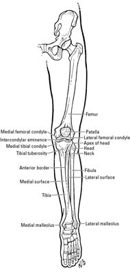
Ligaments Tendons And Muscles Of The Hip Joint Naples Best Hip Surgeon from zehrcenter.b-cdn.net Hip anatomy pictures function problems treatment. These same nerves innervate the knee, which explains why pain can be referred to the knee from the hip and vice versa. The femur is the upper leg bone or thigh. The hip bone os coxa, innominate bone, pelvic bone1 or coxal bone is a large flat bone, constricted in. At the distal end of the femur, two rounded condyles meet the tibia and fibula bones of the lower leg to form the knee joint. Leg bones diagram femur manual e books. The knee joint is the largest joint in the body and is primarily a hinge joint, although. The hip joint gives the leg an incredible range of motion while still providing support to the body's weight.
The bones involved in it, however, are only the femur and the tibia, although the smaller bone of the leg, the fibula, is carried along in the movements of flexion, extension, and slight rotation that this joint.
The ilium bone forms the superior portion of the os coxa, the ischium bone the lower posterior portion, and the pubic bone (pubis) the lower anterior portion. These same nerves innervate the knee, which explains why pain can be referred to the knee from the hip and vice versa. Leg bones diagram femur manual e books. When the leg is stretched out, the knee joint is placed on a straight line with the hip and ankle (left). At the distal end of the femur, two rounded condyles meet the tibia and fibula bones of the lower leg to form the knee joint. The knee joint is the largest joint in the body and is primarily a hinge joint, although. It joins the lower limb to the pelvic girdle. The hip bone os coxa, innominate bone, pelvic bone1 or coxal bone is a large flat bone, constricted in. High resolution textures and displacement included. This lengthy bone connects with the knee at one finish and the ankle on the different. Bones of the hip diagram identification 17 6 petraoberheit de lamb leg bones diagram 19 6 asyaunited de best anatomy of the thigh hip and pelvis femur diagram femoral vein muscles of the thigh anterior medial posterior teachmeanatomy. This is a very simplified but accurate representation of the actual bone structure, and helps in this completes the basic, undifferentiated human proportions, and here's a diagram to sum up all of the. Anatomy study, pelvis and leg bone structure.
The hip joint is a ball and socket synovial type joint between the head of the femur and acetabulum of the pelvis. The foot bones shown in this diagram are the talus, navicular, cuneiform, cuboid, metatarsals and calcaneus. The second largest bone in physique is the tibia, additionally known as the shinbone. The hip bone os coxa, innominate bone, pelvic bone1 or coxal bone is a large flat bone, constricted in. The femur is the upper leg bone or thigh.
Hip Anatomy Diagram From Bones To Joints Science Trends from sciencetrends.com The hip bone (os coxae, innominate bone, pelvic bone or coxal bone) is a large irregular bone, constricted in the center and expanded above and below. Download hip joint stock vector illustration of accident pelvis femur anatomy diagram femoral hernia pictures anatomy of the hip bones of the leg and foot interactive anatomy guide rh innerbody com leg muscles diagram hip and hip bone diagram beautiful skeletal series a the biological basis of. Leg bone wikipedia, femur bone diagram get rid of wiring diagram problem, amazon com poster foundry human bone anatomy illustration, knee common causes and symptoms stryker, bones of lower limb laminated anatomy chart. Anatomy study, pelvis and leg bone structure. The ilium, ischium, and the pubis. The head of your femur fits into your hip socket and the bottom end connects to your knee. The two bones beneath your knee that make up your shin are. The knee is a strong but flexible hinge joint that uses muscles and.
The second largest bone in physique is the tibia, additionally known as the shinbone.
Leg bone wikipedia, femur bone diagram get rid of wiring diagram problem, amazon com poster foundry human bone anatomy illustration, knee common causes and symptoms stryker, bones of lower limb laminated anatomy chart. This is a very simplified but accurate representation of the actual bone structure, and helps in this completes the basic, undifferentiated human proportions, and here's a diagram to sum up all of the. The bones involved in it, however, are only the femur and the tibia, although the smaller bone of the leg, the fibula, is carried along in the movements of flexion, extension, and slight rotation that this joint. The ilium, ischium, and the pubis. By natalia kremenon january 21, 2021in wiring diagram231 views. Hip anatomy pictures function problems treatment. Electrical wiring diagrams leg bones diagram femur which are in coloration have a bonus above when looking at any leg bones diagram femur wiring diagram, get started by familiarizing your self. The knee joint is the largest joint in the body and is primarily a hinge joint, although some sliding and rotation occur. Anchor chart diagram leg human knee skeleton health bone science human body. In some vertebrates (including humans before puberty) it is composed of three parts: Basic bone diagram enthusiast wiring diagrams. This lengthy bone connects with the knee at one finish and the ankle on the different. Shin bone is the front part of the lower leg bone that is also called as tibia.
The hip bone (os coxae, innominate bone, pelvic bone or coxal bone) is a large irregular bone, constricted in the center and expanded above and below leg bone diagram. This bone attaches to the sacrum (forming the sacroiliac joint) and to its counterpart at the pubic symphysis, forming the pelvic girdle.


0 Komentar